


Recently, under the guidance of the team led by Xiangbin Pan from Fuwai Hospital, director Xu Wang and director Shao Zhiyuan and their team at Tongliao People's Hospital successfully completed the first transcatheter aortic valve replacement (TAVR) procedure in the Inner Mongolia Autonomous Region performed entirely under echocardiographic guidance without radiation. This is also the first TAVR procedure in the Tongliao region carried out via the transapical approach.
The procedure was performed without reliance on X-ray fluoroscopy, placing higher demands on the operators' imaging interpretation and procedural coordination. It marks an important breakthrough for Tongliao People's Hospital in minimally invasive interventional treatment of structural heart disease and in the application of ultrasound-guided techniques.
With thorough preoperative evaluation and an individualized treatment plan, the procedure was completed smoothly. Intraoperative hemodynamics remained stable, and postoperative aortic regurgitation was effectively corrected, providing a safe and feasible new minimally invasive treatment option for patients with complex anatomical structures and high risk for conventional open-heart surgery.
Patient Baseline
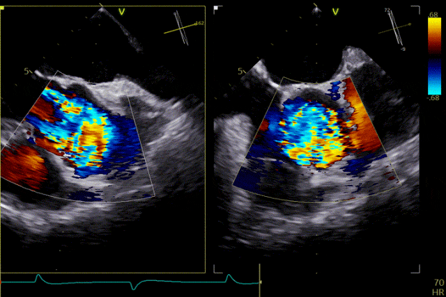
Twenty days ago, he visited the outpatient clinic, where echocardiography indicated aortic valve calcification with severe regurgitation. Recently, the patient again experienced chest tightness and shortness of breath and was admitted for further treatment. His past medical history includes hypertension for 4 years, previous cerebral infarction, and status post cholecystectomy 1 year ago. On admission, the diagnoses were: severe aortic valve insufficiency, grade 3 hypertension (very high risk), and coronary atherosclerotic heart disease.Echocardiography findings showed aortic valve calcification, prolapse of the right coronary cusp with severe insufficiency, increased transvalvular aortic flow velocity, and mild mitral and tricuspid regurgitation. Coronary angiography revealed a normal left main coronary artery, no significant stenosis in the left anterior descending artery, scattered plaques in the circumflex artery, and no significant stenosis in the right coronary artery.
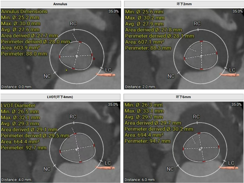
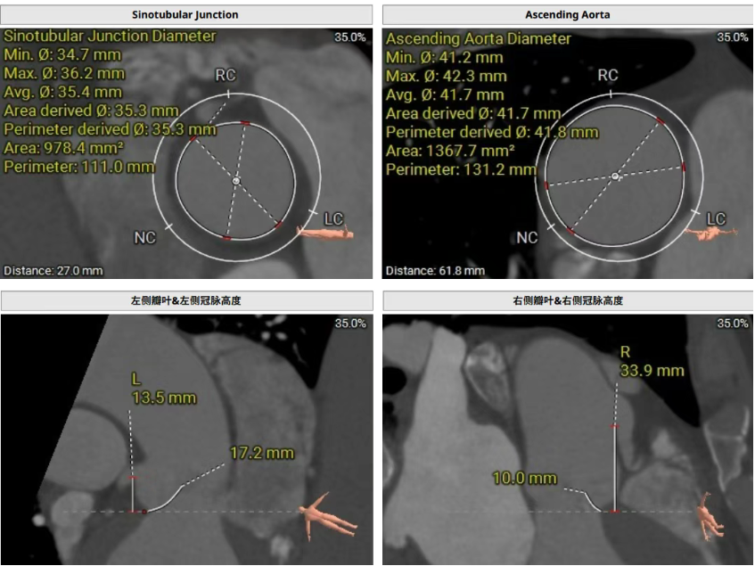
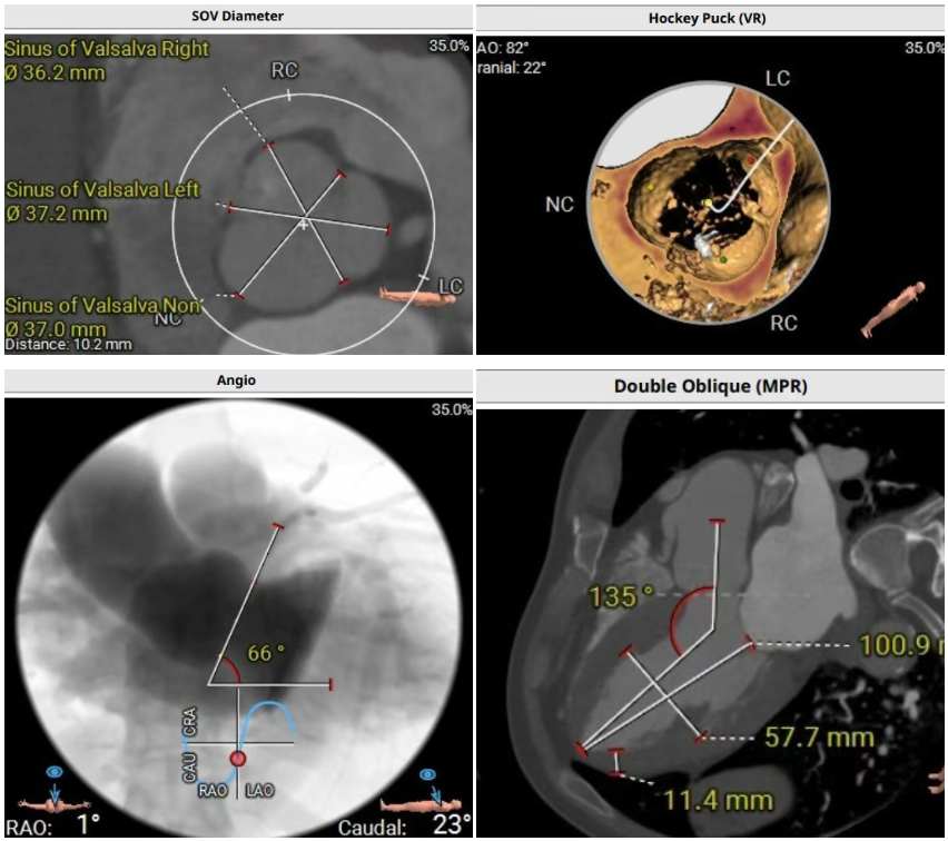
Preoperative Assessment
Preoperative CT evaluation showed that the patient had a tricuspid aortic valve with thickened leaflets. During diastole, prolapse of the right coronary cusp was observed, with mild calcification of that cusp. Three aortic sinuses were present and relatively evenly distributed. The perimeter-derived diameter of the aortic annulus was 28.0 mm. The perimeter-derived diameter of the left ventricular outflow tract (LVOT) was 29.5 mm, with no calcification or stenosis. The sinotubular junction (STJ) measured 35.3 mm in diameter, and the ascending aorta measured 41.8 mm. The coronary ostial heights were as follows: left coronary artery (LCA) 13.5 mm, with a leaflet length of 17.2 mm; right coronary artery (RCA) 33.9 mm, with a leaflet length of 10.0 mm. Mild calcification was noted in the left coronary artery. The patient had a horizontally oriented heart, with an angle of 135° between the left ventricle and the aorta.
Intraoperative Review
Under echocardiographic guidance, the surgical team efficiently and precisely completed valve positioning, deployment, and functional assessment. The procedure proceeded smoothly: engagement of the graspers into the aortic sinuses and positioning were accomplished in one continuous step. The release and anchoring positions of the three graspers were fully consistent with the preoperative assessment, and device manipulation time was approximately 5 minutes. Intraoperative echocardiography showed no paravalvular leak, and hemodynamic parameters remained stable throughout. The procedure was completed successfully without complications.
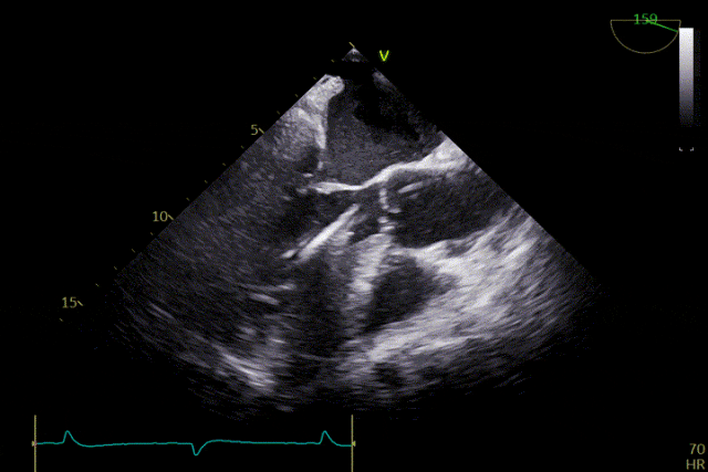
 Preoperative Echocardiography
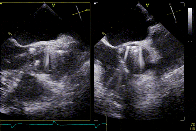
 Delivery system crossing the valve
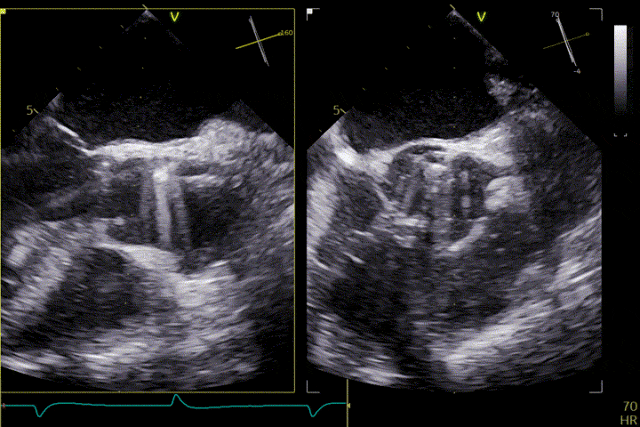
 Deployment of the positioning device
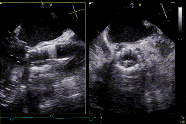
 Advanced into the aortic sinuses
 Valve release
Postoperative Outcome
The patient's severe aortic regurgitation was successfully corrected. Postoperatively, there was no residual regurgitation or paravalvular leak. Postoperative echocardiography showed that the bioprosthetic valve had normal opening and closing function, with a mean transvalvular pressure gradient of 2 mmHg and normal flow velocity. The patient demonstrated good recovery, with a significant improvement in cardiac function compared with the preoperative status.
Summary
Ken-Valve is a TAVR device specifically designed for patients with aortic regurgitation (with or without concomitant stenosis). Its multi-size platform and large-size coverage allow it to accommodate annulus diameters ranging from 23 mm to 33 mm, addressing a previously unmet clinical need in patients with large aortic annuli and predominant regurgitation. The device integrates several key technologies. Its integrated grasping and anchoring design provides stable fixation, which is particularly advantageous in anatomies with little or no calcification. The sealing skirt is designed to reduce the incidence of paravalvular leak after implantation. In addition, the bovine pericardial leaflets treated with a proprietary anti-calcification technology demonstrate favorable biocompatibility and mechanical strength, suggesting good long-term durability. The adjustable delivery system further enables flexible navigation through complex access routes and ensures accurate valve deployment.
The successful completion of the first fully echocardiography-guided, radiation-free transcatheter aortic valve intervention for aortic regurgitation at Tongliao People's Hospital not only provides a safe and effective minimally invasive treatment option for patients with complex aortic valve regurgitation, but also marks a significant advancement in the hospital's capabilities in structural heart disease intervention and ultrasound-guided techniques.
In conventional cardiovascular interventions, reliance on fluoroscopy and contrast agents poses challenges such as iatrogenic injury risk, high technical barriers, limited real-time anatomical visualization, and dependence on large imaging systems. To address these limitations, the structural heart disease team at Fuwai Hospital has spent years developing a fully ultrasound-guided interventional system centered on "visualization, positioning, and directionality" — the PAN technique. The application of the PAN technique reduces radiation exposure and contrast-related risks, and shifts interventional procedures from equipment-intensive and highly dependent models toward a more streamlined and widely accessible approach. Through workflow optimization and technological integration, this method enables complex interventions without large imaging equipment, offering a new pathway to improve procedural safety, expand accessibility, and strengthen capacity at the primary care level.
Disclaimer
The content disclosed in this article is intended for academic exchange purposes only and should not, under any circumstances, be construed as medical advice. Our company makes no representations or warranties regarding the accuracy, completeness, or timeliness of any original, reproduced, or shared content.
The data referenced herein is for informational purposes only and shall not be regarded as advice, recommendations, offers, or invitations. This material is based on publicly available information, internally developed data, and information obtained from other sources deemed reliable. However, no guarantee is made as to the absolute reliability of such information. All opinions and views expressed are based on judgments as of the date of writing and are subject to change at any time without notice.
With respect to any medical devices mentioned in this article, our company makes no representations or warranties regarding their performance or clinical outcomes in diagnostic or therapeutic applications.
- Prev:CSI Frankfurt 2026 | Live LuX-Valve Plus Procedure: Sneak Preview
- Next:A global first! Jenscare Scientific's iJensRobo robotic system successfully completes the first clinical study of the LuX-Valve Plus TTVR system, ushering in a new era of precision intervention for structural heart disease


Privacy Law Copyright © Jenscare Scientific Co., Ltd. All rights reserved. 网站备案/许可证号:浙ICP备17016094号

